Muscle Chart Back - Male Shoulder And Chest Muscles Labeled Chart On White Stock Photo Download Image Now Istock
Muscle Chart Back - Male Shoulder And Chest Muscles Labeled Chart On White Stock Photo Download Image Now Istock. Skeletal muscle human anatomy nerve anatomy leg anatomy body anatomy anatomy back anatomy images human back. Muscle charts of the human body for your reference value these charts show the major superficial and deep muscles of the human body. The most common type of back pain is muscle pain—also called muscle strain or soft tissue strain. Superficial back muscles, intermediate back muscles and intrinsic back muscles.the intrinsic muscles are named as such because their embryological development begins in the back, oppose to the superficial and intermediate back muscles which develop elsewhere and are therefore classed as extrinsic muscles. Nerves in your lower back.
ads/bitcoin1.txt
Five pairs of lumbar spinal nerves labeled l1 to l5 branch off your spinal cord and exit through small holes between the vertebrae. We think this is the most useful anatomy picture that you need. On this page, you'll learn about each of these muscles, their locations and functional anatomy. Certain back muscles extend to other areas, like the shoulders, upper arms, and thighs. When back development is the goal, stick to one of these variations.
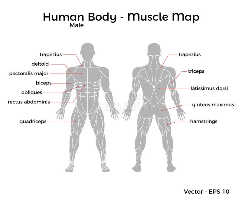
Muscles Chart Description Muscular Body Man Stock Vector Illustration Of Deltoids Health 90796905 from thumbs.dreamstime.com Leaning back to straight vertical and all points in between. We've created a free trigger point chart, which includes fybromyalgia treatment and reflexology information. Facebook twitter google+ linkedin stumbleupon tumblr pinterest reddit vkontakte share via email print. A back muscle that pulls the arm down and back. Your clients will thank you for it! Certain back muscles extend to other areas, like the shoulders, upper arms, and thighs. This diagram shows which muscles in the lower back may be causing you pain. Other muscles are small and cover much less space.
Related posts of back muscles chart muscle recovery anatomy.
ads/bitcoin2.txt
It also helps in extension and lateral flexion of the lumbar spine. Other muscles are small and cover much less space. This increases blood flow to the muscle normalizing it and bringing it back to a healthy state. Symptoms of muscle pain include: The back's muscles start at the top of the back (named the cervical vertebrae) and go to the tailbone (also named the coccyx). Some of these muscles are quite large and cover broad areas. The two trapezius muscles extend from the backbone and base of the skull, across the back and shoulders to join the scapula and the clavicle. This diagram shows which muscles in the lower back may be causing you pain. For more anatomy content please follow us and visit our website: Both the deltoid and the trapezius are firmly attached to the spine of the scapula. There are three different muscle groups found in the back: The trapezius and latissimus dorsi muscles connect the upper limb to the vertebral column. Superficial back muscles, intermediate back muscles and intrinsic back muscles.the intrinsic muscles are named as such because their embryological development begins in the back, oppose to the superficial and intermediate back muscles which develop elsewhere and are therefore classed as extrinsic muscles.
The first step to understanding what your back muscle spasm is telling you is to visit a doctor and get an accurate diagnosis. The most common type of back pain is muscle pain—also called muscle strain or soft tissue strain. There are three different muscle groups found in the back: Others, like sumo deadlifts, have been shown in emg studies—and in the trenches—to focus more on other muscle groups than the back. It is responsible for extension,adduction, and (medial) internal rotation of the shoulder joint.
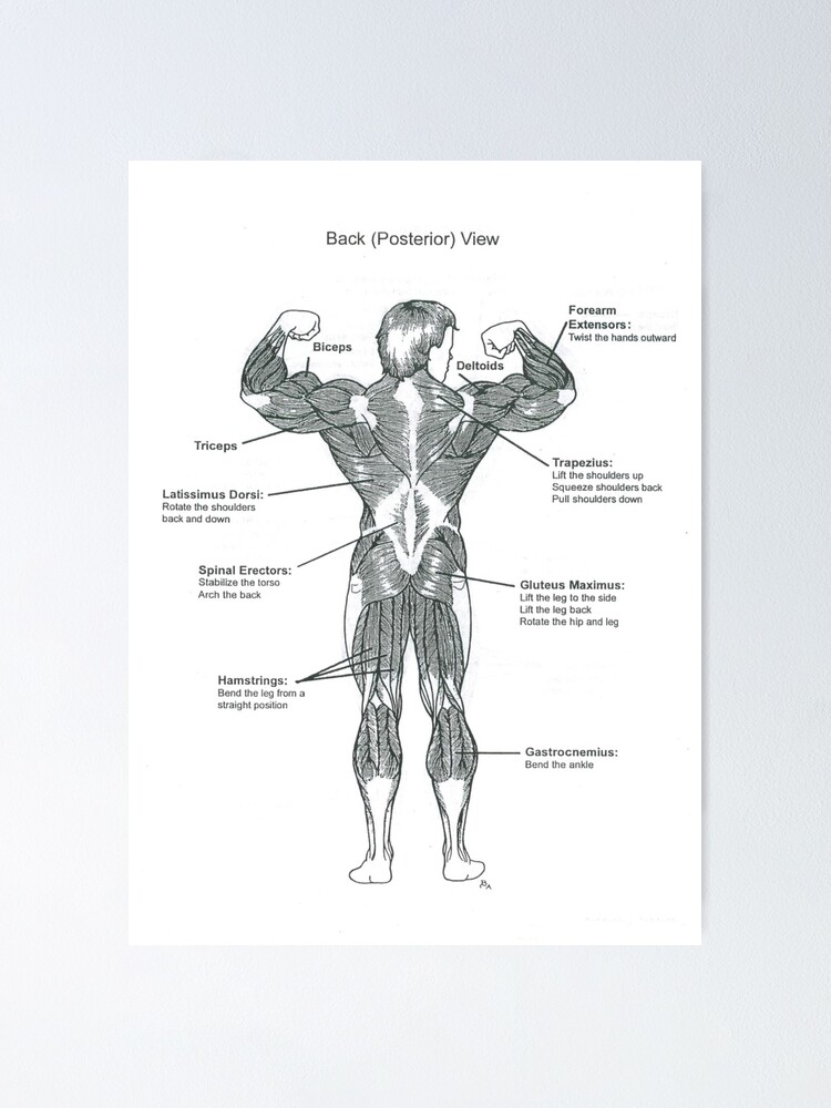
Anatomy Diagram Muscle Chart Back Poster By Superfitstuff Redbubble from ih1.redbubble.net They lift and tilt head and lift or steady the shoulders. The trapezius and latissimus dorsi muscles connect the upper limb to the vertebral column. In general, most back muscle spasms occur because of the following reasons: Both the deltoid and the trapezius are firmly attached to the spine of the scapula. A strain can be an injury to a tendon attachment from muscle to bone. Back pain treatment many individuals will not need extensive treatment for back pain. This diagram shows which muscles in the lower back may be causing you pain. Some of these muscles are quite large and cover broad areas.
Nerves in your lower back.
ads/bitcoin2.txt
Certain back muscles extend to other areas, like the shoulders, upper arms, and thighs. Some of these muscles are quite large and cover broad areas. Both the deltoid and the trapezius are firmly attached to the spine of the scapula. A strain can be an injury to a tendon attachment from muscle to bone. The back's muscles start at the top of the back (named the cervical vertebrae) and go to the tailbone (also named the coccyx). The back anatomy includes the latissimus dorsi, trapezius, erector spinae, rhomboid, and the teres major. Related posts of back muscles chart muscle recovery anatomy. Symptoms of muscle pain include: We've created a free trigger point chart, which includes fybromyalgia treatment and reflexology information. Claim your free copy of the client back care guide today. Others, like sumo deadlifts, have been shown in emg studies—and in the trenches—to focus more on other muscle groups than the back. The part of the nerve that emerges out of the spine is called the nerve root. Superficial back muscles, intermediate back muscles and intrinsic back muscles.the intrinsic muscles are named as such because their embryological development begins in the back, oppose to the superficial and intermediate back muscles which develop elsewhere and are therefore classed as extrinsic muscles.
Nerves in your lower back. In general, most back muscle spasms occur because of the following reasons: For more anatomy content please follow us and visit our website: The two trapezius muscles extend from the backbone and base of the skull, across the back and shoulders to join the scapula and the clavicle. It is responsible for extension,adduction, and (medial) internal rotation of the shoulder joint.
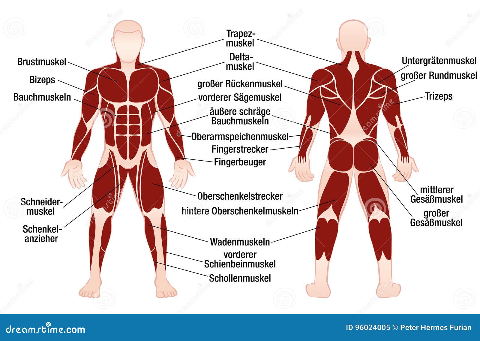
Muscles German Names Chart Muscular Male Body Stock Vector Illustration Of Muscular Bodybuilder 96024005 from thumbs.dreamstime.com Five pairs of lumbar spinal nerves labeled l1 to l5 branch off your spinal cord and exit through small holes between the vertebrae. Other muscles are small and cover much less space. Muscle recovery anatomy 12 photos of the muscle recovery anatomy anatomy of muscle recovery, muscle recovery anatomy, human muscles, anatomy of muscle recovery, muscle recovery anatomy. Postural and active movement muscle, used to tilt and turn the head and neck, shrug, steady the shoulders, and twist the arms. Some of these muscles are quite large and cover broad areas. Related posts of back muscles chart muscle recovery anatomy. We've created a free trigger point chart, which includes fybromyalgia treatment and reflexology information. To see a muscular system picture from the anterior (front) view click here.
It also helps in extension and lateral flexion of the lumbar spine.
ads/bitcoin2.txt
For more anatomy content please follow us and visit our website: It is responsible for extension,adduction, and (medial) internal rotation of the shoulder joint. Facebook twitter google+ linkedin stumbleupon tumblr pinterest reddit vkontakte share via email print. Skeletal muscle human anatomy nerve anatomy leg anatomy body anatomy anatomy back anatomy images human back. Symptoms of muscle pain include: The deltoid, teres major, teres minor, infraspinatus, supraspinatus (not shown) and subscapularis muscles (not shown) all extend from the scapula to the humerus and act on the shoulder joint. To download your free copy click the link. Superficial back muscles, intermediate back muscles and intrinsic back muscles.the intrinsic muscles are named as such because their embryological development begins in the back, oppose to the superficial and intermediate back muscles which develop elsewhere and are therefore classed as extrinsic muscles. The back anatomy includes the latissimus dorsi, trapezius, erector spinae, rhomboid, and the teres major. The first step to understanding what your back muscle spasm is telling you is to visit a doctor and get an accurate diagnosis. Five pairs of lumbar spinal nerves labeled l1 to l5 branch off your spinal cord and exit through small holes between the vertebrae. These structures work together to support the body, enable a range of movements, and send messages from the brain to. A large muscle group in the shoulder, neck and upper back that pulls the head and shoulders backward.
ads/bitcoin3.txt
ads/bitcoin4.txt
ads/bitcoin5.txt
0 Response to "Muscle Chart Back - Male Shoulder And Chest Muscles Labeled Chart On White Stock Photo Download Image Now Istock"

0 Response to "Muscle Chart Back - Male Shoulder And Chest Muscles Labeled Chart On White Stock Photo Download Image Now Istock"
Post a Comment